Restricted Access
This website is intended strictly for qualified research professionals. All products are sold for in-vitro laboratory research use only and are not intended for human or veterinary use.
Statements have not been evaluated by the U.S. Food and Drug Administration. Products are not intended to diagnose, treat, cure, or prevent any disease. Janera Science is not a 503A compounding pharmacy or 503B outsourcing facility.


Date
Category
MOTS-c chemistry sits apart from every other peptide in a research catalogue for one structural reason: the sequence is not written in the nuclear genome. It is written inside mitochondrial DNA.
For decades, the mitochondrial genome was understood as a small, specialized gene set coding for components of the respiratory machinery and nothing else. The identification of a functional peptide sequence inside the 12S ribosomal RNA gene changed that picture.
This article covers what MOTS-c is at the molecular level, how its unusual encoding works, what published laboratory research has examined, and what its sequence means for analytical verification.
This content is provided for informational and educational purposes only and does not constitute medical, pharmaceutical, or legal advice. The materials discussed are intended for laboratory research purposes only and are not for human or animal consumption. They are not intended to diagnose, treat, cure, or prevent any disease.

What Mitochondrial-Derived Peptides Are
Mitochondrial-derived peptides (MDPs) are short peptides encoded by small open reading frames located inside mitochondrial DNA genes, rather than inside the nuclear genome where almost all known signalling peptides originate.
The mammalian mitochondrial genome encodes 13 messenger RNAs, 22 transfer RNAs, and 2 ribosomal RNAs, the 12S and 16S subunits (International Journal of Molecular Sciences, 2022). Every identified MDP is transcribed from a short open reading frame sitting inside one of those two rRNA transcripts.
Three MDP groups are described in the literature:
Humanin, encoded within 16S rRNA and first reported in 2001
Small humanin-like peptides (SHLPs), also encoded within 16S rRNA
MOTS-c, encoded within 12S rRNA and first reported in 2015
The naming follows the location. MOTS-c stands for mitochondrial open reading frame of the 12S rRNA type-c.
Why the Encoding Location Matters Chemically
Peptides derived from a larger parent protein are common in research supply. Fragment-derived compounds such as the thymosin beta-4 fragment are cleaved or synthesized from a known nuclear-encoded protein sequence.
MOTS-c is a different case. The sequence overlaps a functional ribosomal RNA gene, meaning the same stretch of mitochondrial DNA carries structural rRNA information and a peptide-coding reading frame at the same time.
MOTS-c Structure and Sequence
MOTS-c is a 16-amino-acid peptide with the sequence MRWQEMGYIFYPRKLR (Cell Metabolism, 2015).
Written in three-letter code, the chain runs from the N-terminus to the C-terminus as follows:
H-Met-Arg-Trp-Gln-Glu-Met-Gly-Tyr-Ile-Phe-Tyr-Pro-Arg-Lys-Leu-Arg-OH
The molecular formula is C101H152N28O22S2 (PubChem, 2026). Calculated from that formula, the average molecular mass is approximately 2,174.6 g/mol for the free peptide.
Property | Value |
|---|---|
Length | 16 residues |
One-letter sequence | MRWQEMGYIFYPRKLR |
Molecular formula | C101H152N28O22S2 |
Average molecular mass | ~2,174.6 g/mol |
Genomic origin | 12S rRNA gene (MT-RNR1), mitochondrial DNA |
Reading frame length | 51 base pairs |
Physical form (research supply) | Lyophilized powder |
Composition Notes Worth Recording
Three features of the sequence carry practical weight for anyone characterizing a batch.
Two methionine residues sit at positions 1 and 6. Methionine is among the most oxidation-prone residues in peptide chemistry.
One tryptophan sits at position 3, and two tyrosines sit at positions 8 and 11. Tryptophan is also oxidation-sensitive, and the aromatic residues together give the peptide strong ultraviolet absorbance at 280 nm.
Four basic residues appear across the chain: arginine at positions 2, 13, and 16, plus lysine at position 14. The strongly basic C-terminal region drives the net positive charge of the peptide at neutral pH.

The 12S rRNA Encoding Story
The original identification came from an in silico screen rather than from protein isolation. Investigators searched the human 12S rRNA sequence for potential short open reading frames and found one of 51 base pairs carrying a strong Kozak sequence, translating to 16 amino acids (Cell Metabolism, 2015).
Translation Happens Outside the Mitochondrion
Here is the detail that most summaries skip.
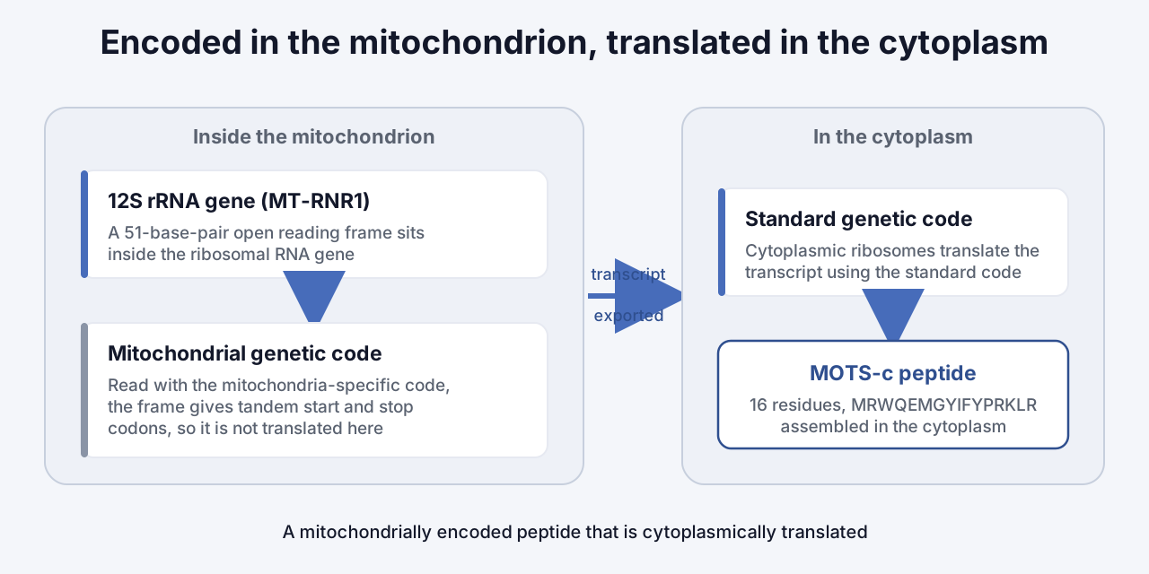
Mitochondria run their own genetic code, which differs from the standard code used in the cytoplasm. Read using the mitochondria-specific code, the MOTS-c reading frame produces tandem start and stop codons and does not yield the peptide.
Translation of MOTS-c therefore occurs in the cytoplasm using the standard genetic code, which requires the transcript to be exported from the mitochondrion first (Cell Metabolism, 2015). The peptide is mitochondrially encoded but not mitochondrially translated.
Sequence Conservation
The MOTS-c sequence, and particularly the first 11 residues, is highly conserved across 14 species including humans and mice (Frontiers in Endocrinology, 2023). Conservation across that evolutionary distance is one of the standard signals researchers use to argue that a reading frame is functional rather than incidental.
What Published Laboratory Research Has Examined
The following summarizes observations recorded in laboratory models. None of it describes outcomes in people outside the specific measurements noted, and none of it establishes any use beyond laboratory research.
The 2015 Discovery Work
The original report described a cellular mechanism in which MOTS-c inhibits the folate cycle and the de novo purine biosynthesis pathway tethered to it. That inhibition raises intracellular AICAR, which in turn activates AMP-activated protein kinase (AMPK) (Cell Metabolism, 2015).
Skeletal muscle was identified as the primary tissue in which these effects were recorded. The same study reported differences in body weight and glucose tolerance between treated and untreated mice maintained on a high-fat diet (Cell Metabolism, 2015).
Nuclear Translocation Under Metabolic Stress
A 2018 study examined where the peptide goes inside a cell under stress conditions. Under basal conditions, MOTS-c was detected predominantly in association with mitochondria, with a small nuclear pool.
Glucose restriction, serum deprivation, and oxidative stress agents each transiently increased nuclear translocation, and chromatin-associated MOTS-c rose significantly within one hour of glucose restriction (Cell Metabolism, 2018). Translocation required AMPK activity.
Once in the nucleus, MOTS-c was found to interact with NRF2, a stress-responsive transcription factor, and to associate with antioxidant response elements in target genes (Cell Metabolism, 2018).
This is the finding that made MOTS-c structurally interesting to a broader field. It documented a factor encoded in the mitochondrial genome acting on nuclear gene expression, a direction of communication that had not previously been demonstrated.
Endogenous Levels and Physical Activity
A 2021 study measured endogenous MOTS-c in human skeletal muscle biopsies and reported an approximately 11.9-fold increase in relative levels following exercise, with levels remaining elevated after a four-hour rest (Nature Communications, 2021).
Note what this measurement is. It records the concentration of a naturally occurring peptide in tissue, sampled before and after an activity. It is an observation about endogenous physiology, not a finding about any administered material.
The K14Q Variant: A Single-Residue Structure-Function Case
This is the part of MOTS-c chemistry with the clearest structural teaching value, and it is rarely covered well.
A naturally occurring mitochondrial DNA polymorphism, m.1382A>C, changes the fourteenth residue of the peptide from lysine to glutamine, producing the variant known as K14Q MOTS-c (Aging Cell, 2015).
One residue. A basic side chain swapped for a neutral amide side chain.

What the Substitution Does in Laboratory Assays
A 2024 study identified protein kinase CK2 as a direct binding partner of MOTS-c and characterized the interaction using dot blot, cell-free kinase activity, and surface plasmon resonance assays (iScience, 2024).
Against that binding target, the K14Q variant showed roughly 16-fold lower binding affinity for the CK2 alpha subunit than the reference sequence, and did not activate CK2 in the cell-free assay (Diabetes, 2024).
Feature | Reference MOTS-c | K14Q variant |
|---|---|---|
Residue 14 | Lysine (basic) | Glutamine (neutral amide) |
Source | Reference mitochondrial sequence | m.1382A>C polymorphism |
CK2 alpha binding affinity | Reference | ~16-fold lower |
CK2 activation, cell-free assay | Recorded | Not recorded |
Why This Matters for Sequence Verification
A single-residue difference in a 16-residue chain shifts the mass by a small amount and changes the charge state, and it materially changes behaviour in a binding assay.
That is the practical argument for sequence-level confirmation rather than purity figures alone. A chromatographic purity number describes how much of the sample is a single species. It does not, on its own, confirm which species. Identity confirmation by HPLC and mass spectrometry is what closes that gap.
Analytical Considerations Specific to This Sequence
MOTS-c is produced for research supply by solid-phase peptide synthesis, the same route used across the research peptide field. Its composition raises two points worth checking on batch documentation.
Oxidation products. With two methionines and a tryptophan in a 16-residue chain, methionine sulfoxide and tryptophan oxidation products are the impurity classes most worth confirming were screened. Each carries a characteristic mass shift that mass spectrometry resolves.
Counter-ion and net peptide content. With three arginines and one lysine, the peptide carries a substantial positive charge and correspondingly binds counter-ions from the synthesis and purification process. Gross mass is not net peptide mass, and the difference is not trivial for a sequence this basic.
Material is supplied as a lyophilized powder for this reason among others. Removing water from a peptide containing oxidation-sensitive residues is the standard approach to limiting degradation over a storage window.
Frequently Asked Questions
What is MOTS-c?
MOTS-c is a 16-amino-acid peptide encoded by a short open reading frame within the 12S ribosomal RNA gene of mitochondrial DNA, with the sequence MRWQEMGYIFYPRKLR. It was first reported in 2015 and belongs to a group called mitochondrial-derived peptides (Cell Metabolism, 2015).
What does the name MOTS-c stand for?
MOTS-c is a contraction of mitochondrial open reading frame of the 12S rRNA type-c. The name identifies where in the mitochondrial genome the reading frame sits rather than describing any property of the peptide.
How is MOTS-c different from other research peptides?
Almost all signalling peptides are encoded in the nuclear genome. MOTS-c is encoded inside mitochondrial DNA, though it is translated in the cytoplasm using the standard genetic code because the mitochondria-specific code yields tandem start and stop codons for this reading frame (Cell Metabolism, 2015).
What is the molecular weight of MOTS-c?
The molecular formula is C101H152N28O22S2 (PubChem, 2026), giving a calculated average molecular mass of approximately 2,174.6 g/mol for the free peptide. Salt forms carry additional mass from the counter-ion.
What is the K14Q MOTS-c variant?
K14Q MOTS-c is a naturally occurring sequence variant arising from the m.1382A>C mitochondrial DNA polymorphism, which replaces the lysine at position 14 with glutamine (Aging Cell, 2015). In laboratory binding assays the variant showed approximately 16-fold lower affinity for the CK2 alpha subunit than the reference sequence (Diabetes, 2024).
Which residues in MOTS-c are most sensitive to oxidation?
The two methionine residues at positions 1 and 6 and the tryptophan at position 3 are the oxidation-sensitive positions. Mass spectrometry resolves the resulting oxidation products by their characteristic mass shifts.
What does research use only mean for a material like this?
Research use only indicates that a material is supplied exclusively for laboratory research and is not approved or intended for human or animal use. A fuller explanation is available on the research use only page.
Key Takeaways
MOTS-c is a 16-amino-acid peptide, sequence MRWQEMGYIFYPRKLR, encoded by a 51-base-pair open reading frame inside the mitochondrial 12S rRNA gene (Cell Metabolism, 2015).
The peptide is mitochondrially encoded but cytoplasmically translated, because the mitochondria-specific genetic code yields tandem start and stop codons for this reading frame (Cell Metabolism, 2015).
A 2018 study documented MOTS-c translocating to the nucleus under metabolic stress in an AMPK-dependent manner, where it associated with NRF2 and antioxidant response elements (Cell Metabolism, 2018).
A single lysine-to-glutamine substitution at position 14 reduced binding affinity for the CK2 alpha subunit by roughly 16-fold in laboratory assays, which is why sequence identity confirmation matters alongside purity figures (Diabetes, 2024).
Two methionines and one tryptophan make oxidation products the impurity class most worth confirming on batch documentation for this particular sequence.
All materials supplied by Janera Science are for laboratory research purposes only. They are not for human or animal consumption and are not intended to diagnose, treat, cure, or prevent any disease. Purchasers are responsible for confirming that intended use complies with all applicable regulations. See the frequently asked questions page for ordering and documentation details.
Every batch verified
Janera Science publishes third-party Certificates of Analysis for its research peptides. To review HPLC purity, heavy metal, endotoxin and sterility results for current lots, visit our lab results page. For the regulatory context behind how these materials are supplied, see our overview of what Research Use Only means.